Services
Dental treatment plan
The treatment usually begins with professional consultation referred by the dentist responsible for basic dental care. If considerable time has passed since the basic care or if the problem is acute, the treatment begins with a comprehensive oral examination. In both cases, the condition of your whole dentition is examined and a long-term prognosis and function of your teeth are assessed. The necessary radiographs, like the panoramic radiographs of the dentition and jaw area, as well as the more specific images, are taken in this phase at the latest. In the case of extensive occlusal and esthetic rehabilitations, optical impressions and facial images are taken for planning the treatment and smile design. Bite Reconstruction
We prepare a clear treatment plan and give a cost estimate for each treatment. The final cost estimate may vary by up to 10 % from the one given. Once you have approved the treatment plan, we can agree on starting and scheduling the treatment. On average, the treatment begins 1–2 months after the plan has been made and approved.
Diagnostic wax-up
Diagnostic wax-up or diagnostic CAD refers to the 3D design made for partial esthetic care or a full-mouth restorative bite treatment. The design guides the production of the final structures.
In the 3D design, the anatomy, occlusal load distribution, phonetics, and esthetics of your dentition in proportion to your lips and face are optimized. The design is converted into physical dental cast models through 3D printing which enables the production of molds based on these models. With the help of molds, the design is tested before producing the final structures. Treatments using bite reconstruction, veneer, and injection molding techniques require diagnostic wax-up.
1st visit
- Bite registration
- Teeth scanning
- 3D face scanning or photography
30 min
2nd visit
- Testing prepared wax-up on teeth
- Removal of test wax-up
- Visit in the initial treatment phase or separately
30 min


Implant
A dental implant is a screw made of titanium, and just like the root of your tooth, it works as an anchor in the jawbone. The root of the missing tooth is replaced with an implant, then the visible part of the implant crown is attached to it. A dental implant is particularly suitable for replacing one tooth, but by using several implants an even larger number of missing teeth can be replaced.
1st visit
- Clinical examination
- Radiography
- Treatment plan
- Teeth scanning
30 min
2nd visit
- Individual care pathway
Teeth straightening with clear aligners
A current practice in today’s dental care, teeth straightening with clear aligners, involves using a series of thin transparent plastic foils. The method is used for treating small and moderate malocclusions that are traditionally treated with solid dental braces. Teeth straightening with clear aligners is planned using CAD based on optical dental impressions and photographs, taking your wishes into account. We discuss the preliminary computer-designed treatment plan with you, and make the necessary changes. The completed plan indicates the duration of your straightening treatment, the number of visits needed, and the estimated costs. With the help of CAD, the forces and trajectories applied for teeth movement are optimized, which makes the straightening procedure quicker compared to traditional treatment with dental braces. It also reduces the number of your visits to the dentist. In most cases, both the upper and the lower dental arches are treated.
During the treatment process, you change the sets of aligners to new ones every week or every two weeks, depending on the system. Each mouthpiece is thin and flexible, and it fits the shape of the teeth and moves the teeth in the desired direction. Each set of aligners is worn 20–22 hours a day, and they are removed only when eating and cleaning them and your teeth. During and after the straightening treatment, the teeth always tend to return to their initial position. This can be prevented by wearing an adhesive wire on the inner part of the tooth or with a retainer, which is thicker than a proper straightening aligner, at night.
1st visit
- Clinical examination
- Radiography
- Treatment plan
30 min
2nd visit
- Teeth scanning
- Photography of teeth, bite, and face
30 min
3rd visit
- Approval of treatment plan
- Modification of treatment plan
15 min
4th visit
- Interproximal reduction (IPR)
- Placing attachments
- Starting use of first set of aligners
30–60 min
5th–8th visit
- Follow-up of treatment progress
- Interproximal reduction (IPR), when necessary
15–30 min
9th visit
- Teeth scanning
- Photography of teeth, bite, and face
30 min
10th visit
- Removal of attachments
- Starting use of retainers
- Finishing procedures needed
Overall duration of treatment: individual duration 1–3 years, retention period included


Bite guard
A bite guard is a custom-fit therapeutic teeth protector. It is made of hard acrylic plastic with a 3D milling machine or of resin using 3D printing. Most bite guards are designed to be placed on the upper teeth. The therapeutic property of a bite guard is based on its guiding effect on the jaw. When you bite your teeth together, the guard protects your dentition and guides your jaw into a relaxed position, which reduces muscular symptoms. The guard is worn only at night, but at the beginning of the treatment it can also be worn in the daytime. A good guard is both effective in relieving symptoms and pleasant to wear. It protects not only the teeth but also tooth-bearing structures and jaw joints against masticatory forces.
1st visit
- Teeth and bite scanning
15 min
2nd visit
- Fitting of the finished bite guard
30 min
3rd visit
- Follow-up of bite guard usage
15–30 min
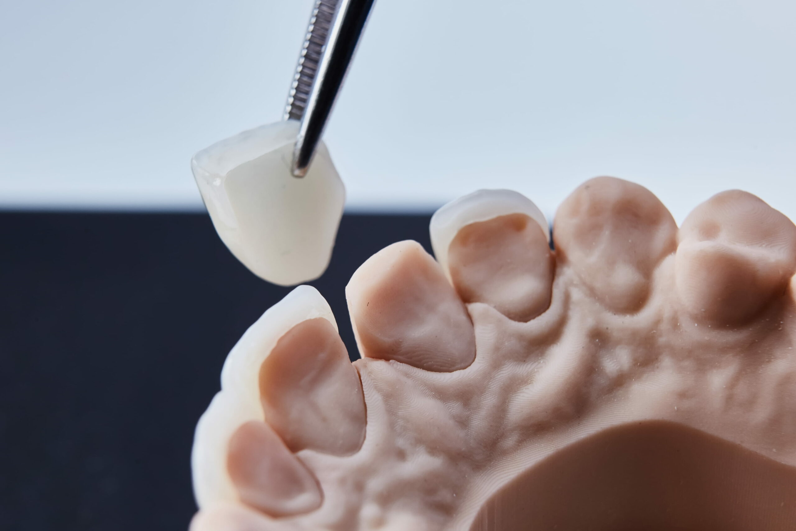
Crown
Teeth with extensive fillings, chips, root canal treatments, and severe wear are treated with a ceramic dental crown. A tooth with a ceramic crown feels and looks like a tooth of your own. It is also extremely durable and protects the remaining tooth tissue by covering it up.
1st visit
- Local anesthesia
- Removal of old fillings
- Teeth preparation
- Teeth scanning
- Placing temporaries
30 min/tooth
2nd visit
- Local anesthesia
- Removal of temporaries
- Crown cementation or bonding
30 min/crown


Crown lengthening
Crown lengthening involves a surgical procedure performed as a preparatory measure to address a severely worn-down or decayed tooth before it is crowned. The gum of the tooth that needs rehabilitation is opened, and 2–4 millimeters of the surrounding bone is removed. After the surgery, the gum will heal over a couple of weeks and settle down during the next 3–6 months. When the gum has settled down, the permanent crown can be placed on the exposed, healthy tooth tissue.
1st visit
- Clinical examination
- Radiography
- Treatment plan
30 min
2nd visit
- Teeth scaling, cleaning gum pockets, when necessary
45 min
3rd visit
- Dental surgery
60–90 min
4th visit
- Removal of sutures
15 min
5th visit
- Crown treatment 3–6 months after the surgery
Veneer
The incisors (front teeth) are often treated with dental veneers for esthetic reasons. Malpositioned and discolored teeth under treatment can be masked with ceramic veneers. These enable rapid and natural changes without long-term straightening treatments. They also provide an excellent means of closing gaps between teeth and lengthening worn-down teeth. Good veneers are natural in esthetics and they blend in with your other teeth. Only a very minimal amount of drilling is needed for the veneers and therefore, veneers are not suitable for treating severe malposition of teeth. In these cases straightening treatment combined with teeth whitening gives the best result. If the form, length or the shade of your teeth needs altering, the veneers can be installed at the end of the teeth straightening treatment, also.
1st visit
- Clinical examination
- 3D face scanning
- Teeth scanning
30 min
2nd visit
- Fitting of wax-up
15 min
3rd visit
- Teeth preparation
- Teeth scanning
- Placing temporaries
45–60 min
4th visit
- Bonding veneers
45–60 min

Injection molding technique
When using the injection molding technique, fillings are made by injecting filling material through transparent silicon molds. Shaping your fillings is unnecessary except for the last finishing touch because the shape of the tooth emerges as the silicon mold made from the diagnostic wax-up is filled with the injectable filling material. The final shape of your teeth will correspond exactly to the diagnostic wax-up.
The technique is ideally suited to the anterior teeth area in the cases where it is important to mask malpositioned teeth, to increase the size of teeth, to close open gaps between teeth, and to cover discolored teeth in a tooth-sparing manner. The technique is also appropriate as part of more extensive bite rehabilitation. Plastic veneers manufactured with this technique are much less costly and quicker to make than their ceramic counterparts. In a visual sense, the fillings made using the injection molding technique are highly esthetic.
1st visit
- Bite registration
- Teeth scanning
- 3D face scanning
30 min
2nd visit
- Testing prepared wax-up on teeth
- Removal of test wax-up
- Shade selection for teeth
30 min
3rd visit
- Local anesthesia
- Fitting of molds
- Removal of old fillings
- Making new fillings
- Bite check-up
30–120 min
4th visit
- Bite check-up
- Polishing of fillings
30 min
In an extensive project, the third visit is divided into several shorter visits.
Bridge
The purpose of a dental bridge is to replace a missing tooth or teeth with the support of adjacent or remaining teeth. If the adjacent teeth to your missing tooth are completely flawless, there is no need to make a bridge. Therefore, a bridge is best suited to situations where the teeth next to the missing tooth have been fixed, one way or another.
1st visit
- Local anesthesia
- Teeth preparation
- Teeth scanning
- Placing temporaries
45–75 min
2nd visit
- Local anesthesia
- Removal of temporaries
- Attaching the bridge
45–60 min /bridge
Preparatory work before the first visit, when necessary
30–60 min


Partial cover restorations
Glass ceramic is an excellent way to restore your molars and premolars (that is, back teeth and the teeth between the back teeth and canine teeth). As a structure, ceramic inlays, onlays and overlays are hard, sustainable and esthetic. The bond of the ceramic restorative material to the tooth is excellent, and due to its hardness and adhesiveness, the bond weakens at a much slower rate than a corresponding bond between a composite filling and a tooth. Owing to a strong adhesive bond, the ceramic structure integrates into your tooth, strengthening the remaining edges of your teeth by binding them together.
1st visit
- Local anesthesia
- Removal of old fillings
- Teeth preparation
- Teeth scanning
- Placing temporaries
30 min /tooth
2nd visit
- Local anesthesia
- Removal of temporaries
- Restoration bonding
30 min /tooth
Wisdom teeth removal surgery
Wisdom teeth are the third set of molars that only seldom have enough room to erupt in modern man’s dentition. It is very common that the gum surrounding a wisdom tooth is infected by a partially erupted tooth or the adjacent tooth in front of it starts decaying. Even if your wisdom tooth has erupted properly, cleaning it can be difficult. Wisdom tooth removal by surgery is a completely painless procedure, apart from the unpleasant feeling caused by local anesthesia. Therefore, removing these teeth as a preventive measure at an early age is recommended since the procedure is easiest to perform then. In the surgery, a cut is made next to the wisdom tooth on the side of the cheek. The bone is usually drilled from the side of the cheek and the top of the tooth so that the tooth and its roots can be cut up and removed. The wound is closed tightly with sutures.
The risks inherent in the operation are individual, and they depend on the location of the wisdom tooth and the tooth socket nerve. However, in experienced hands, the surgery can be considered almost risk-free. In situations involving a risk of nerve injury, 3D radiography provides valuable information on the location of the nerve in relation to the wisdom tooth. This makes it possible to perform the extraction procedure according to a detailed plan and without injuring the nerve.
1st visit
- Clinical examination
- Radiography
- Treatment plan
30 min
2nd visit
- Preparations
- Local anesthesia
- Surgery
- Post-operative instructions
45–60 min


Botulinum toxin therapy
Botulinum toxin (Botox) is used as symptomatic and preventive treatment to paralyze hyperactive and sore muscles. The substance is administered by injection into the muscle under treatment, and it blocks the access of the nerve impulses from the nerve into the muscle. The targeted muscles will be paralyzed only partially. They will maintain their functional ability, but the maximum force they can produce will decrease. As regards a hyperactive and enlarged muscle, the desired effect is achieved because the injection returns its masticatory force to the normal level.
The paralysis that results from the injection is temporary and will last about 3–6 months. Due to the paralysis of the muscle, the mass of it will decrease. Therefore, those having unnecessarily large and strong masticatory muscles as a result of bruxism benefit from the injections, even after the paralyzing effect.
Botulinum toxin is a widely investigated and safe drug with only marginal side effects. Pregnancy, breastfeeding, and a medical condition that involves muscular dystrophy are reasons a patient may not receive Botulinum toxin therapy. Our clinic provides Botulinum toxin therapies to treat migraines, bruxism and for esthetic care of the face and neckline.
A bite guard does not eliminate bruxism—neither does Botulinum toxin—but the appropriate co-administration of these can offer effective control of the masticatory symptoms and protect bite. Botulinum toxin therapy to treat bruxism is recommended if any of the following occur:
- a bite guard is not an option
- the response of the bite guard is inadequate
- patient’s bite breaks the bite guards
1st visit
- Bite examination
- Bite guard check-up
- Treatment plan
- Prescription
30 min
2nd visit
- Botulinum toxin injections
- Post-operative instructions
15 min
3rd phase: Patient contacts by phone/email
- Assessment of effect
Two weeks from the injection
4th phase: Patient contacts by phone/email
- Assessment of effect
- Need for further injections
- Prescription writing
3–12 months from the injection
5th visit
- Botulinum toxin injections
- Post-operative instructions
15 min
Second opinion
I will provide a second opinion on extensive prosthetic treatment plans and masticatory disorders that do not respond to treatment.
In masticatory disorders, an overall examination of bite is necessary. In addition to the mastication muscles, it involves the muscles in the head and neck area, and the jaw joints. Moreover, we will examine your current bite guard (its bite, thickness, type and fit), if you have one, and determine if it is still effective and beneficial. I will express my view on the previous treatment plan, review the radiographs, and confirm/disprove the diagnoses made earlier. During the visit, we will make a plan for treatment, draw up a cost estimate, and explain, clearly and understandably, the causes and consequences of the disorder to you.
In the case of extensive prosthetic treatments, such as bite reconstruction, I will present the alternative treatment plans and submit my own view and cost estimate on the treatment plans already made. Together, we will discuss the advantages and disadvantages of all the treatments possible for your situation. I will confirm the previous diagnoses and comment on the timing of the treatments.
A second opinion is beneficial when:
- you have an extensive treatment plan.
- you have received dental treatment but the disorder continues.
- you are unsure whether treatment is necessary, or you worry about its costs.
A second opinion is beneficial because:
- you will get an independent opinion from an experienced specialist.
- your understanding of the issue will increase.
- you will get more alternatives.
- you will get a competing offer for the same treatment.
Before getting a second opinion:
- ask your dentist for your dental records and dental chart.
- ask your dentist for your dental radiographs for the past three years.
- prepare for your visit by thinking about the following questions:
- What is the diagnosis in the treatment plan?
- What are the alternative treatments?
- How do these treatments improve my dental health?
- What are the risks involved in the treatments?
- What are the costs?
- What are the consequences without treatment?
- What treatments do I need in the future?
1st visit
- Clinical examination
- Diagnosis
- Treatment plan
- Cost estimate
30 min

Bite reconstruction
Bite reconstruction involves full-mouth occlusal rehabilitation that makes the collapsed teeth feel and look like they had never needed any treatment at all.
